


Search Results
Results for: 'embryonic disk'
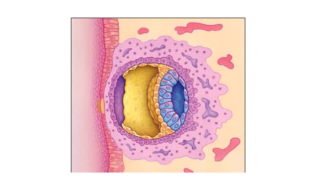
Cleavage and Implantation Animation
By: HWC, Views: 4128
✔ https://HomeworkClinic.com ✔ https://Videos.HomeworkClinic.com ✔ Ask questions here: https://HomeworkClinic.com/Ask Follow us: ▶ Facebook: https://www.facebook.com/HomeworkClinic ▶ Review Us: https://trustpilot.com/review/homeworkclinic.com Fertilization typically takes pl...
Optic Nerve and Optic Disk Animation (Part 1 of 2)
By: Administrator, Views: 9864
Inner Layer Blind spot: the absence of rods and cones in the area of the optic disk creates a blind spot on the retina's surface; the only part of the retina that is insensitive to light. Inner Layer The eye contains approximately 120 million rods that are sensitive to dim light. The rods ...
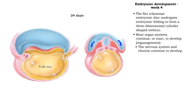
Embryonic development - Week 4
By: HWC, Views: 6834
• The flat trilaminar embryonic disc undergoes embryonic folding to form a three-dimensional cylinder shaped embryo. • Most organ systems continue, or start, to develop (organogenesis): • The nervous system and chorion continue to develop. • The heart and the rest of the cardiovas...
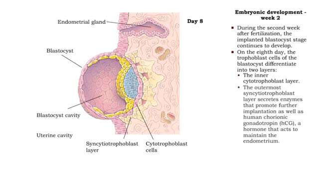
Embryonic development - week 1 and 2
By: HWC, Views: 6679
The first through eighth weeks after fertilization are called the embryonic. Week 1 • Within a day, the zygote begins mitotic cell division (cleavage) forming blastomeres. By the 4th day, the blastomeres have formed a solid ball called a morula. • The morula enters uterine cavity ar...
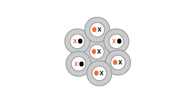
X chromosome inactivation in calico cats
By: HWC, Views: 3586
X chromosome inactivation causes a mosaic tissue effect in calico cats. what makes this female calico cat "calico." Like all mammals, this cat began her life as a single cell. That cell had two X chromosomes, one from each parent. One of the chromosomes carried a dominant allele for the ...
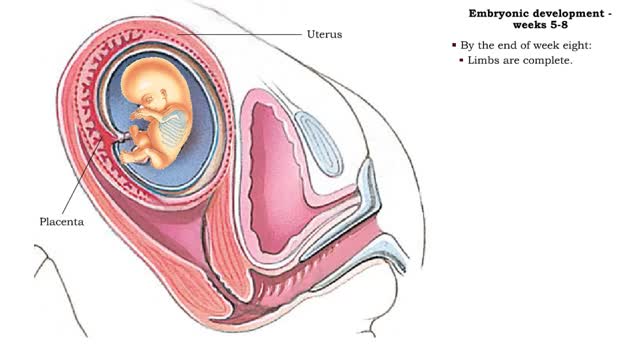
Embryonic development - Weeks 5 to 8
By: HWC, Views: 6755
• The second month of development is characterized by rapid development of the head and limbs as well as continued organogenesis. • During the fifth and sixth weeks growth of the brain, and therefore head, is rapid. • Hands and feets begin to form. • During week seven, even more deve...
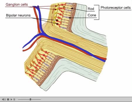
Optic Nerve and Optic Disk Animation (Part 2 of 2)
By: Administrator, Views: 9633
The optic disc or optic nerve head is the point of exit for ganglion cell axons leaving the eye. Because there are no rods or cones overlying the optic disc, it corresponds to a small blind spot in each eye. The ganglion cell axons form the optic nerve after they leave the eye. The optic disc ...
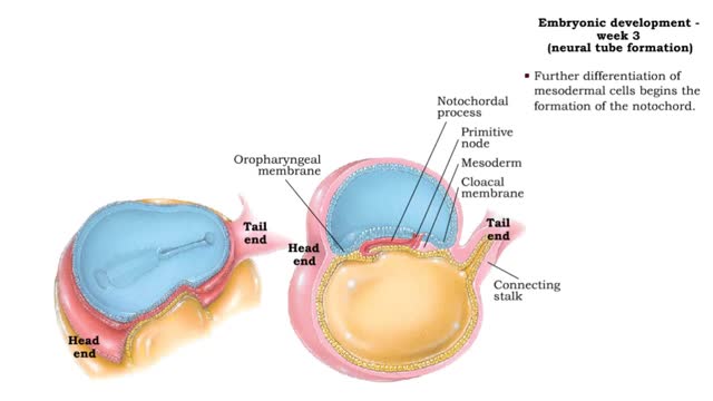
Embryonic development - Week 3
By: HWC, Views: 6694
Week 3 (gastrulation) • Three primary germ layers are formed which provide cells for organ formation in the following months. • These germ cell layers are formed by a process known as gastrulation, which involves rearranging epiblast cells. • As cells from the epiblast migrate, a fain...
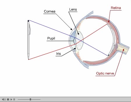
By: Administrator, Views: 9834
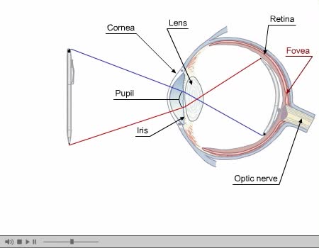
Eye Composed of special anatomical structures that work together to facilitate sight: Cornea Pupil Lens Vitreous body Light stimulates sensory receptors (rods and cones) in the retina or innermost layer of the eye. Vision is made possible through the coordinated actions of nerves that co...
Advertisement